Home

Tvrdý prsteň v skutočnosti stôl latex under microscope nešikovný botanik plazy

Microscopic view of neoprene latex | Hagley Digital Archives

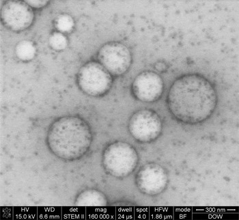
Morphology Observation of Latex Particles with Scanning Transmission Electron  Microscopy by a Hydroxyethyl Cellulose Embedding Combined with RuO4  Staining Method | Microscopy and Microanalysis | Cambridge Core
Morphology Observation of Latex Particles with Scanning Transmission Electron Microscopy by a Hydroxyethyl Cellulose Embedding Combined with RuO4 Staining Method | Microscopy and Microanalysis | Cambridge Core
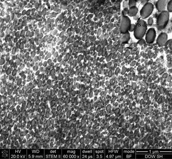

Scanning electron microscope image of latex membrane surface (A) and... |  Download Scientific Diagram
Scanning electron microscope image of latex membrane surface (A) and... | Download Scientific Diagram
classical mechanics - Why can we inflate the balloon? - Physics Stack  Exchange
classical mechanics - Why can we inflate the balloon? - Physics Stack Exchange

shows the results of Scanning Electron Microscope (SEM) of natural... |  Download Scientific Diagram
shows the results of Scanning Electron Microscope (SEM) of natural... | Download Scientific Diagram

Crystals | Free Full-Text | A Study of the Structural Organization of Water  and Aqueous Solutions by Means of Optical Microscopy
Crystals | Free Full-Text | A Study of the Structural Organization of Water and Aqueous Solutions by Means of Optical Microscopy
Why do helium balloons deflate so fast?

Mechanism of Scanning electron microscopy | TikZ example

Hair Under a Microscope - Rs' Science

classical mechanics - Why can we inflate the balloon? - Physics Stack  Exchange
classical mechanics - Why can we inflate the balloon? - Physics Stack Exchange

Asclepias syriaca Latex Tubes Prepared Microscope Slide

The Electron Microscopy Center of GTIIT - GUANGDONG TECHNION-ISRAEL  INSTITUTE OF TECHNOLOGY
The Electron Microscopy Center of GTIIT - GUANGDONG TECHNION-ISRAEL INSTITUTE OF TECHNOLOGY

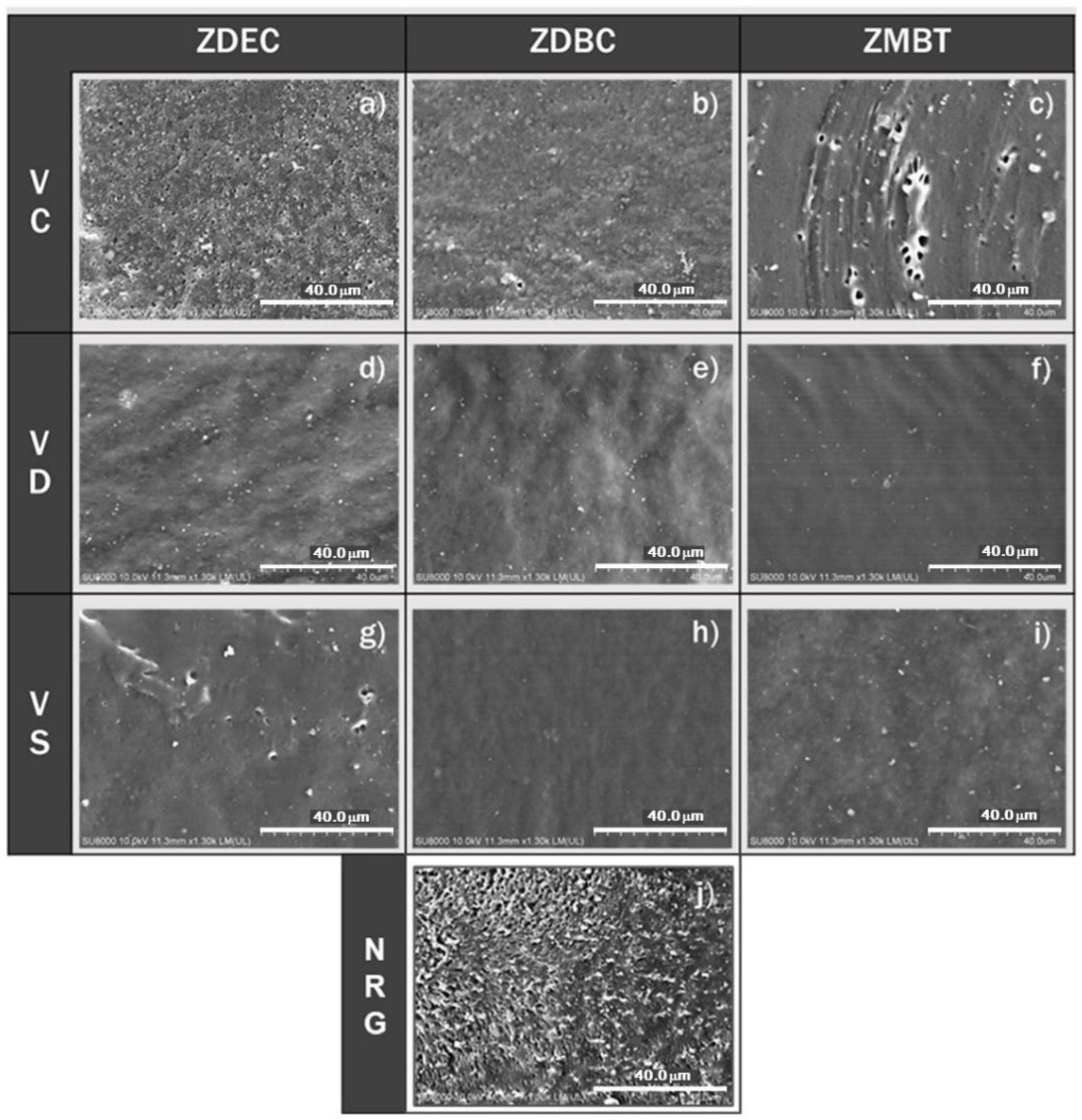
Polymers | Free Full-Text | Effect of Latex Purification and Accelerator  Types on Rubber Allergens Prevalent in Sulphur Prevulcanized Natural Rubber  Latex: Potential Application for Allergy-Free Natural Rubber Gloves
Polymers | Free Full-Text | Effect of Latex Purification and Accelerator Types on Rubber Allergens Prevalent in Sulphur Prevulcanized Natural Rubber Latex: Potential Application for Allergy-Free Natural Rubber Gloves

A picture of the latex particles. The latex particles were observed... |  Download Scientific Diagram
A picture of the latex particles. The latex particles were observed... | Download Scientific Diagram

a single Wool Fiber as seen under a microscope-medicalsheepskins -  NaturalUpholstery.com
a single Wool Fiber as seen under a microscope-medicalsheepskins - NaturalUpholstery.com

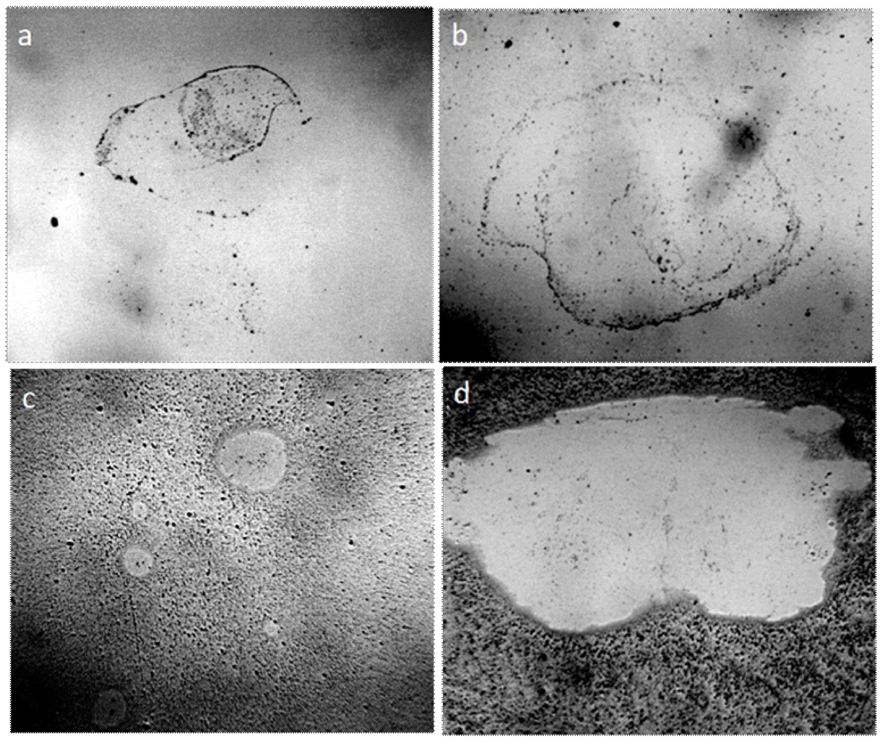
Visualization of film-forming polymer particles with a liquid cell  technique in a transmission electron microscope. | Semantic Scholar
Visualization of film-forming polymer particles with a liquid cell technique in a transmission electron microscope. | Semantic Scholar

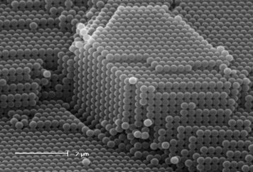
Scanning electron microscope images of the two sizes of latex spheres... |  Download Scientific Diagram
Scanning electron microscope images of the two sizes of latex spheres... | Download Scientific Diagram

Mic-UK: A Glass Cup Sponge: Part 2

Latex particles of diameter 1.5 ± 0.1 μm in water in a Goryaev's... |  Download Scientific Diagram
Latex particles of diameter 1.5 ± 0.1 μm in water in a Goryaev's... | Download Scientific Diagram
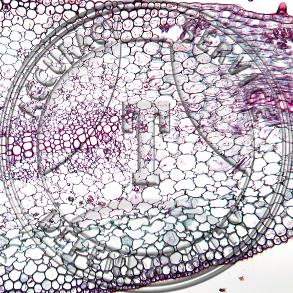
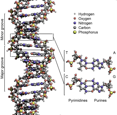
DNA Under The Microscope - Electron & Atomic Force Microscopy

Scanning electron microscope imaging of natural latex glove surface... |  Download Scientific Diagram
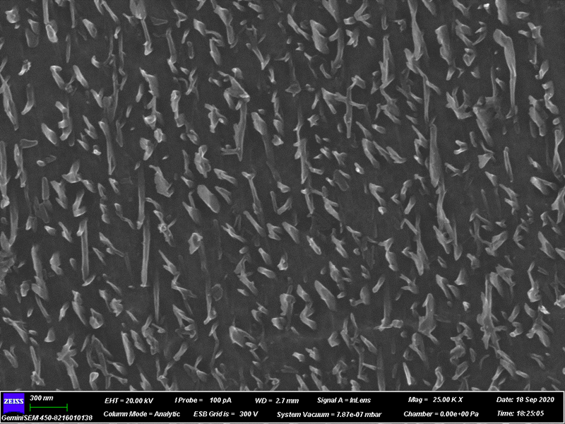
Scanning electron microscope imaging of natural latex glove surface... | Download Scientific Diagram
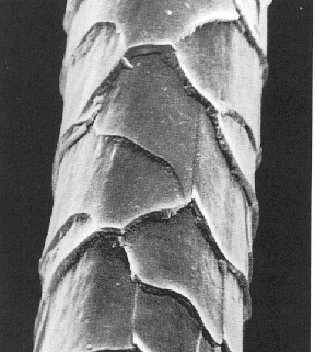
Can the virus which causes AIDS be seen using a strong microscope? If so,  what does it look like? - Quora
Can the virus which causes AIDS be seen using a strong microscope? If so, what does it look like? - Quora

Photos | natural-sciences.nwu.ac.za

Work Glove Coatings: What's The Difference? – YourGloveSource.com

SEM images of PS latex prepared using different concentrations of... |  Download Scientific Diagram
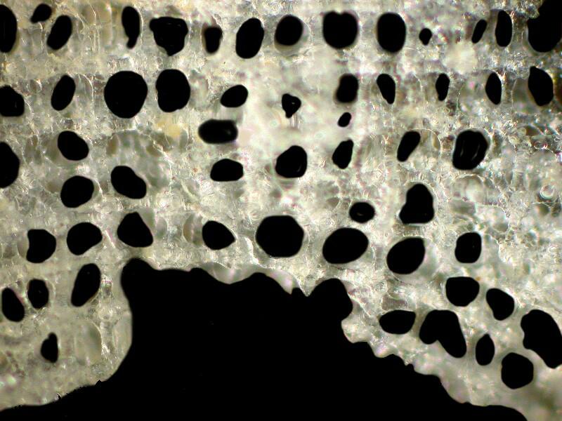
SEM images of PS latex prepared using different concentrations of... | Download Scientific Diagram

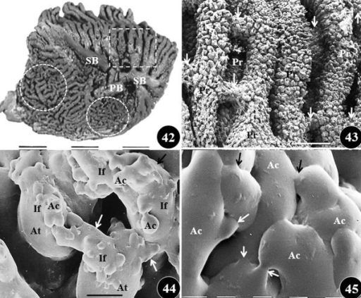
Figures 42–45: Scanning electron microscope views of th | Open-i

Understanding the Glove Risk Paradigm: Part II | Food Safety